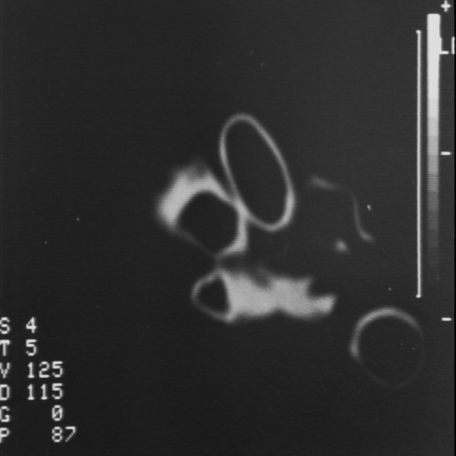
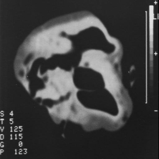
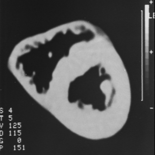
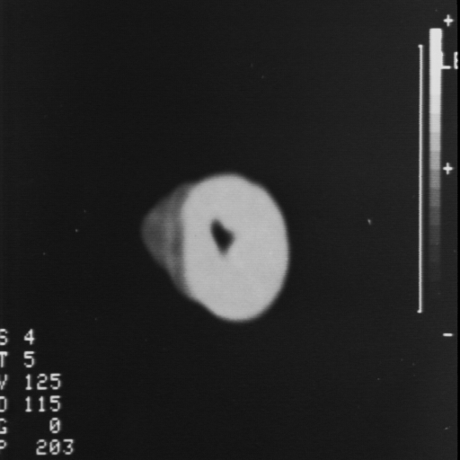
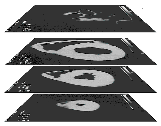
Modelling of the Human Heart
To get a spatial 3-D model of the human
heart, 32 computer-tomography slices
of the human heart were generated. These slices were positioned
equidistantly in a Z-direction which represented the axis originating
from the apex toward the base. The resolution on the X-Y plane was
512 x 512 points and was determined by a scanning procedure.
Because of the integrity of the model, it was necessary to
guarantee the exact overlaping of all slices. Before digitalisation,
two reference points on every picture were marked in order to adjust
all data.
3-D model of the human heart:
 |
The precision of this model was later further reduced to meet the
requirements of the simulation.