Figure 1. Anatomical picture of the human heart.
| Previous: Introduction | Top | Next: Cross-section Preparation |
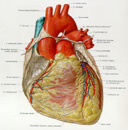
The heart is the central organ of the blood-vascular system and consists mainly of a hollow muscle of conical form that pumps blood into a set of outgoing vessels - arteries and through them to all parts of the body [8]. The blood is re-collected in the veins and returned to the heart closing the loop of the circulation of the blood. The heart is positioned obliquely in the chest and placed between the lungs. The broad end, called base, is directed upwards, backwards and to the right. All the great vessels are attached to the heart at its base. The opposite end, called the apex, is directed downwards, forward and to the left. The human heart is divided into two halves by a septum. Each half, the left and the right, is further divided into an upper atrium and a lower ventricle. The left half of the heart pumps the blood through the body. From the cavity of the left ventricle, the blood is carried into the aorta, and further through smaller arteries to the periphery. The blood flows from the body through the peripheral veins, and finally through the superior and inferior vena cava into the right atrium. This flow is termed the greater or systemic circulation. From the right atrium, the blood is passed into the right ventricle and from there into the truncus pulmonalis and further through the pulmonary arteries into the lungs. From there it is returned through the pulmonary veins into the left atrium. This flow is termed the smaller or pulmonary circulation. The blood is passed further into the left ventricle, so that both circulations are connected.
There are four heart valves regulating the blood flow in the heart. Two are between each atrium and ventricle and are called atrioventricular valves. The valve in the left half is also called the mitral valve and the one in the right half, the tricuspid valve. The third valve, positioned at the beginning of aorta, is called the aortic valve, and the fourth, at the beginning of the truncus pulmonalis, the pulmonary valve. They all prevent blood flowing in wrong direction.
The heart wall, which surrounds all four heart chambers, is composed of three layers. The inner layer, the endocardium, is very thin and covers all the inner surface of the heart. It is continued in the arteries as the tunica intima. The middle layer is the myocardium, the muscle that actually performs pumping function. The muscle fibres of this layer are gathered in several layers running in different directions. The muscle fibres for the atria and the ventricles are separated. The outer layer is called the epicardium. It, too, consists of two layers. The inner, visceral one is the serous membrane. It is tightly attached to the myocardium and is very rich in adipose tissue. At the heart base, near the great vessels, it is folded back as a fibrous, outer layer, which is also known as the pericardium. It is much stronger and surrounds the whole heart as a pericardial sac. Both layers are divided by a capillary space filled with serous fluid. The wall of the great vessels originating from heart base has a similar structure. The inner part, the tunica intima, is a continuation of the endocardium. The middle part, the tunica media or tunica muscularis, consists of muscular fibers. The outer layer, the tunica adventitia is fibrous tissue, similar to the pericardium.
The heart has double circulation, both functional and nutritional. The functional circulation consists of all heart chambers and the great vessels of the heart. These parts serve for conducting the blood through the heart. The nutritional circulation brings blood to all parts of the heart, because the heart, as with every organ in the human body, needs the delivery of oxygen and nutrients. There are several anatomical variations, but, in general, the two main arteries, the left and right coronary arteries originate from the aorta, just above the aortic valve. The right coronary artery runs backwards, between the truncus pulmonalis and the right atrium to the posterior surface and in the sulcus interventricularis posterior, just between the left and right ventricle, to the apex where its small branches anastomose with branches from the left coronary artery. The left coronary artery runs forwards onto the anterior surface of the heart. It divides then into two arteries. The left anterior descending artery runs into the sulcus atrioventricularis anterior, just between left and right ventricle, to the apex, where its small branches anastomose with the right coronary artery. The second branch, the left circumflex artery, runs around the left margin of the heart on the posterior surface. The veins of the heart follow the coronary arteries at the beginning, but then join together on the posterior surface as the sinus coronarius, which ends in the right atrium.
Figure 1. Anatomical picture of the human heart.
| Previous: Introduction | Top | Next: Cross-section Preparation |