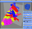
Figures 8, 9.Typical 3-D editor environment. Selected portion of the 3-D model and three 2-D editing windows.
| Previous: Practical Experience with Heart Tissue Distinction | Top | Next: Results |
All mentioned mistakes in the generated spatial heart model must be corrected, either manually or automatically, by a computer program. It is very difficult to imagine the failures in the space or to see their relationship to other tissues in the space. It was expected that a kind of 3-D editing would be of great help in improving the spatial heart model. The primary goal in designing the spatial editor was to allow the user to easily spot and correct the smaller failures in the generated 3-D models. This model was built from processed VHD cross-sections in such a manner that each pixel represented a cube in 3-D space.
In order to minimise the number of objects, the cubes that are fully obscured have been extracted and eliminated in the graphical presentation. Additionally, identical neighbouring cubes have been represented as a single larger object. The number of objects still remains too large to allow a smooth interactive work and rendering of the model; therefore, the redisplaying of the 3-D model is done only upon user request.
The editor can display a 3-D model of all six distinct tissues or any subset of them in a user-selectable portion of the heart. The selected portion can be rotated and zoomed in and out in order to note all the desired details and possible mistakes.
Figures 8, 9.Typical 3-D editor environment. Selected portion of the 3-D model and three 2-D editing windows.
A model cross-section is user selectable and distinguishably coloured in the 3-D view. Mistakes can be corrected on a set of two-dimensional windows that allow the simultaneous display of the three consecutive cross-sections: previous, selected and next. The user can draw on the selected cross-section with brushes of various sizes in any of the six tissue colours, which correspond to adding/removing portions of a tissue to/from the cross-section. The position of the actual brush is shown in all three sections, demonstrating how the change on the selected one will affect the preceding and succeeding cross-sections. Immediately after a change is made in the 2-D window, it is also applied to the 3-D model, keeping it consistent with actual changes on the selected cross-section.
There are two additional functions that can be selected at start-up that automatically modify the 3-D model. The user can select the reduction of the model size by a factor n, taking portions of the input model made of n´ n´ n pixels and replacing them with a single pixel in the output model, representing the tissue most common in the reduced portion. After the reduction, the input model is backed up and replaced with the reduced one. The second function can find eventual small openings or discontinuities in the selected tissue (exposing other tissues) and repair them by adding a small part of missing tissue onto the corresponding cross-sections. In this way the small holes in the pericardium are automatically filled.
| Previous: Practical Experience with Heart Tissue Distinction | Top | Next: Results |