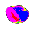
Figure 4. X-Y cross-section 1450 after tissue determination in the resolution of 512 x 512 pixels. Pericardium - black, myocardium - pink, arterial blood - red, venous blood - blue, coronary vessels - green, fat - yellow.
| Previous: Cross-sections Preparation | Top | Next: Spatial Editor |
The most important and, in quantity, the most represented tissues in the heart are the myocardium, the adipose tissue and the pericardium. The greatest volume is also represented by the heart chambers and the coronary vessels, which are normally filled with blood. To enable the computer to distinguish between these main different tissues, they were painted with six different Windows system colours, including the surrounding space that was coloured white.
A set of photographs of X-Y thorax cross-sections from the VHD was used to begin with. Adobe Photoshop 4.0 was used for the basic graphic manipulation of the slices. The pericardium was determined first, and painted black. Selection was made with the very useful wand tool, which selects all neighbouring pixels with similar colours. It was noticed from the photographs that it is practically impossible to distinguish between the epicardium and pericardium; therefore, we do not distinguish these two tissues. Then everything around the pericardium not belonging to the heart was painted white. Next, all the heart chambers were painted in a similar way; the left atrium and ventricle with red and the right atrium and ventricle with blue. They were all quite dark on the photographs and easily distinguished from neighbouring structures. The left ventricle, usually much larger in the working heart, is quite small in our model and looks more like a fissure. The endocardium, which normally lines the entire interior, is microscopically thin, so it was not painted. The wall of the great vessels originating in the heart chambers, was also painted black, like the pericardium. The myocardium was clearly separable from other tissues and painted brown, including all the different layers of muscle tissue that are difficult to distinguish. Finally, the adipose tissue with the coronary vessels remained. The adipose tissue was white-yellow on the photographs and the blood vessels dark spots of different sizes in it. It was coloured yellow, and the coronary arteries, green. Sometimes it was quite difficult to decide where to draw the border between the two tissues because of similar pixel colour. In such cases the borders were drawn subjectively. The described procedure was implemented on each of the 156 cross-sections, from the heart apex to the base. Cross-section 1450 as it appeared after processing, is given in Figure 4, and an AVI animation of all the cross-sections can be viewed.
Figure 4. X-Y cross-section 1450 after tissue determination in the resolution of 512 x 512 pixels. Pericardium - black,
myocardium - pink, arterial blood - red, venous blood - blue, coronary vessels - green, fat - yellow.
The resulting painted cross-sections were rescanned in a resolution similar to that in the Z direction, which was around 1mm, and put together into a 3-D model of the heart. Now all the mistakes that were made in the 2-D editing of the cross-sections could be seen. The main problems were the pulmonary vessels at the base of the heart. Some parts of them, usually the upper and the lower, were missing. This happened because in the beginning they were painted only if they were connected with the heart chambers. On some earlier or later cross-sections they were omitted. Also, the pericardium was discontinued in some places, particularly on the posterior surface of the heart. These defects were also clearly seen on the Y-Z cross-sections that were derived from the 3-D model in order to help correct the mistakes.
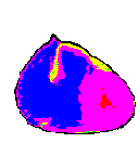
Figure 5. Generated X-Z cross-section 50 in the resolution of 146 x 151 cubes. Pericardium - black, myocardium -
pink, arterial blood - red, venous blood - blue, coronary vessels - green, fat - yellow.
The Y-Z cross-sections have been edited already, thus the mistakes can rarely be seen (Figure 6). The pericardium is closed and other tissues are also connected.
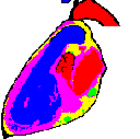
Figure 6. Generated Y-Z cross-section 75 in the resolution of 152 x 151 cubes. There are no mistakes left in tissue
determination.
The next problem arose with the smaller coronary vessels. The left and right coronary arteries, the circumflex artery and the largest veins are visible and one can follow such vessels from their origin to the periphery. But there were some discontinuations of the smaller vessels that did not have a uniform course in the 3-D view. There are also some artifacts, such as spots painted on some cross-sections as vessels, but actually not corresponding to vessel tissues. They look like a single pixel or group of pixels apart from the coronary vessels. The resulting 3-D model is given in Figure 7.
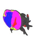
Figure 7. Cross-section of the 3-D model of the human heart. Pericardium - black, myocardium - pink, arterial blood -
red, venous blood - blue, coronary vessels - green, fat - yellow.
| Previous: Cross-sections Preparation | Top | Next: Spatial Editor |